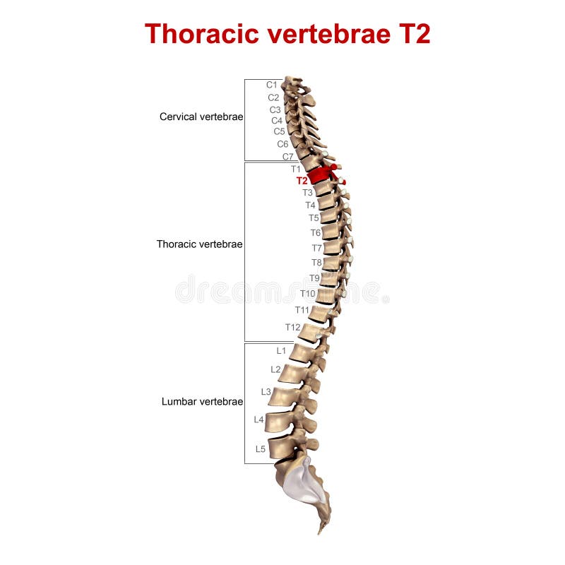
MRI (Magnetic Resonance Imaging) provides a detailed view of the spine’s anatomy, offering critical insights for diagnosing and treating spinal disorders. This comprehensive guide explores various aspects and techniques of spinal MRI, including T1 and T2-weighted images, thoracic spinal fusion, and laminectomy procedures.
Axial MRI of the Lumbar Spine focuses on T2-weighted images at the L4 level, highlighting structures like the cauda equina, surrounded by cerebrospinal fluid (CSF), which appears bright on these images. This view also effectively visualizes the neural foramina, posterior bony elements, and paraspinal muscles, offering a clear picture of the spinal anatomy.
Normal Mid-Sagittal MRI scans of the Lumbar Spine, presented in both T1 and T2-weighted images, provide a comprehensive view of the lumbar spine. These scans encompass all five lumbar vertebrae, the sacrum, and the lower thoracic vertebrae, allowing for a detailed examination of the spine’s structure and potential abnormalities.
T2 (transverse relaxation time) is a crucial parameter in MRI, indicating the rate at which excited protons reach equilibrium or lose phase coherence. This measurement is vital in determining the spinal anatomy and detecting any anomalies.
Thoracic Spinal Fusion, a surgical procedure involving the placement of screws and rods to stabilize the spine, is necessary in cases of instability due to injury, deformity, or pain. This surgery and its aftercare are crucial for patients’ recovery and long-term spinal health.
A thoracic laminectomy, another surgical procedure, involves removing the lamina portion of vertebrae to access and remove tumors or alleviate pressure on the spinal cord. This surgery may include adding instrumentation to stabilize the vertebrae post-operation.
Spine Disorders, such as herniated discs, are well-diagnosed through MRI imaging. The vertebral column, comprising 33 vertebrae, can develop various disorders, necessitating detailed imaging for accurate diagnosis and treatment planning.
Endplate impaction fractures and the subsequent surgical interventions, such as anterior diskectomy and fusion, are also well-evaluated using MRI scans. These procedures can lead to immediate symptom improvement for patients.
For further detailed information and visual guides, refer to the MRI Spine Anatomy, Normal Sagittal MRI, MRI Basics, Thoracic Spinal Fusion Guide, Thoracic Laminectomy Procedure, Herniated Disc Information, Spinal Ligamentous Injury MR Imaging, and Thoracic Laminectomy Details for comprehensive insights into spinal MRI imaging and related surgical procedures.


