Spinal nerves are complex structures composed of ventral and dorsal roots. The ventral roots primarily carry motor outflow axons and autonomic fibers. It is common for several adjacent spinal roots to innervate each muscle. Below is a list of specific muscles and their associated nerve roots:
Ankle: S1
Superficial Reflexes:
Corneal reflex (blink reflex): This is an involuntary blinking response triggered by corneal stimulation. The afferent pathway is the nasociliary branch of the ophthalmic branch (V1) of the trigeminal nerve (5th nerve), and the efferent pathway is the facial nerve (7th nerve).
Abdominal reflex: This involves the contraction of superficial abdominal muscles when the abdomen is lightly stroked.
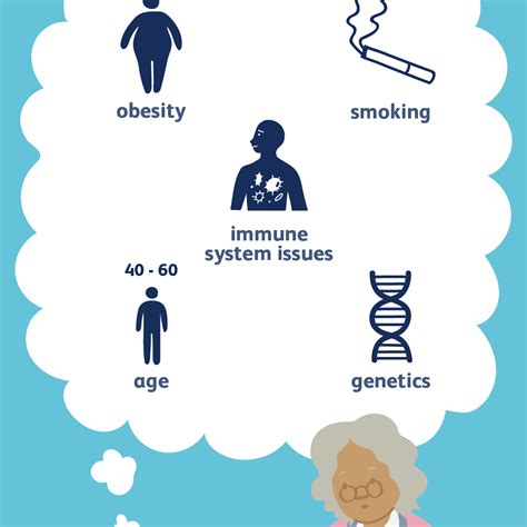
Leg pain that originates in the back is frequently referred to as “sciatica,” though this term specifically pertains to the sciatic nerve. Sciatica describes pain following the route of the S1 nerve root distribution: in the back of the thigh, back of the calf, and foot. According to CreakyJoints, Dena Barsoum, MD, a physiatrist at HSS, explains that the medical term for sciatica is lumbar radiculopathy, often occurring when the S1 nerve root in the low back gets pinched.
Importantly, the major nerve roots to examine for issues are L4, L5, and S1, as they are the most commonly affected. Pain radiating along the posterior thigh and the posterolateral aspect of the leg may be due to an S1 or L5 radiculopathy. S1 irritation may extend pain to the lateral aspect of the foot, while L5 radiculopathy may send pain to the dorsum of the foot and the large toe.
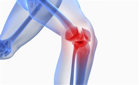
When treating a pinched nerve, it’s crucial to understand that nerves typically branch off the spinal cord through spaces between the vertebrae. If these spaces shrink, they can squeeze the nerve root, causing symptoms in the area served by the nerve.
Using the S1 nerve as an example, it arises from myelomere S1 opposite the T12 vertebra, descends as part of the cauda equina, and emerges from the first sacral foramen. This can be seen in Figure 41-2 A, which illustrates a myelomere of the spinal cord and one of its associated spinal nerves.
Anterior Lumbar Interbody Fusion (ALIF) is considered minimally invasive surgery, utilizing anatomical corridors without major disruption of tissue planes. For those wondering about the size of an ALIF scar, it is very similar to that of a C-section.

For further information and resources, please refer to the following links:
Spinal Nerve Roots
Stanford Medicine 25
Low Back Pain
Arthritis or Sciatica
Neurological Exam
Radiculopathy
Treating a Pinched Nerve
Human Anatomy
ALIF Surgery