The spine’s structure and function play a critical role in overall health and well-being. The normal thoracolumbar spine, which includes both the thoracic and lumbar regions, is relatively straight in the sagittal plane and exhibits a double curve in the coronal plane. Specifically, the thoracic spine curves posteriorly (kyphosis), while the lumbar spine curves anteriorly (lordosis). In a healthy spine, there should be no lateral curvature.
Significant spinal curvatures can lead to various health issues. For instance, large and progressive curves, especially those over 70 degrees, can compress the lungs, causing shortness of breath or quick fatigue. Similarly, progressive thoracolumbar curves can lead to a premature feeling of fullness in the stomach due to hunger satiety.
The spine naturally has three curves: the cervical curve in the neck with a slight forward arch, the thoracic curve in the upper back curving slightly backward, and the lumbar curve in the low back with a slight forward curve. Proper alignment of these curves ensures balance across the spine, shoulders, hips, knees, and ankles, leading to evenly distributed body weight.
In surgical interventions for thoracic curves, which are tethered from the front (anterior), an incision is made on the side of the chest, between the ribs. This approach allows for an unobstructed view of the spine during surgery, with the involved lung being temporarily deflated by the anesthesiologist and reinflated post-procedure.
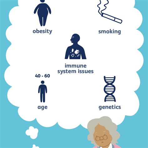
Thoracic hyperkyphosis, or pathologic kyphosis in the thoracic region, is a condition where this natural outward curve becomes excessive. Abnormal curves can occur in both the thoracic and lumbar regions, with severity ranging from minor (as low as 10 degrees) to severe cases (exceeding 100 degrees).
In thoracic spine issues, a fractured vertebral body can lead to spinal cord compression. The thoracic spine is the most common location for osteoporotic fractures, though lumbar fractures are also frequent. Conventional radiographs (X-rays) are typically used as the initial imaging exam for evaluating spinal fractures.
For more detailed information, refer to the resources from University of Washington, Hospital for Special Surgery, Harvard Health, UCSF Health, Columbia Neurosurgery, HSS Kyphosis Overview, Albany Medical Center, Flatback Syndrome at Columbia, and Scoliosis Imaging Overview.